Cystoscopy & Laser Ablation for Vets
Cystoscopy provides a minimally invasive technique for the diagnosis and treatment of ectopic ureters in dogs (as well as the investigation and diagnosis of many other urinary tract issues).
The ectopic ureter(s) can be seen as an abnormal opening in the wall of the urethra.
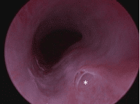
Cystoscopic image of the urethra of female dog with an ectopic ureter. The opening of the ectopic ureter is visible in the foreground.
Once it has been identified a diode laser can be used, via the cystoscope, to remove the wall of the ectopic ureter. The wall is removed back to the level of the bladder neck. This essentially turns the ectopic ureter from a ‘tunnel’ into a ‘trough’ and restores the opening to a more normal anatomic position.
This technique has been associated with a good outcome and has significant advantages over open surgery. The procedure is successful in resolving or improving incontinence. However, affected dogs often have additional urinary abnormalities such as urethral sphincter mechanism incompetence.
One study reported laser ablation of either unilateral or bilateral ectopic ureters in 30 dogs (Berent 2012). Fourteen dogs (47%) were continent without the need for additional treatment. However, with additional treatment in the remaining dogs 23/30 (77%) were ultimately continent.
Reference
Berent AC, Weisse C, Mayhew PD, Todd K, Wright M, Bagley D. (2012) Evaluation of cystoscopicguided laser ablation of intramural ectopic ureters in female dogs. Journal of the American Veterinary Medical Association 240, 716-725 As far as we are aware, we are the only practice in the North East that have experience with this technique.