Elbow Dysplasia
What is elbow dysplasia?
Elbow dysplasia (ED) is a developmental condition of growing dogs. There are several different components to it and different factors that contribute to its occurrence. A dog that is affected by ED will usually show lameness which may affect one or both of the front legs. Any dog can be affected by dysplasia but it is typically seen in larger breeds. Dogs like Labradors appear to be particularly affected.
The lameness associated with ED is not caused by a single problem. Factors that lead to the development of a dysplastic joint involve genetics (though specific genes have not yet been identified), rate of growth, nutrition and environment. This last factor can include the amount of exercise that your puppy gets. Unfortunately development of the condition is not well understood so giving specific recommendations about prevention and management are difficult.
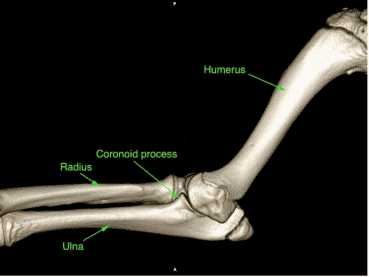
The elbow is the joint formed by the humerus, radius and ulna. Dysplasia results when the bones do not match each other well (known as incongruency). There are various abnormalities but the result of these is overloading at parts of the joint. The most common problem is fragmentation of the medial coronoid process. This lip of bone on the ulna can become detached resulting in a fragment of bone and cartilage, leading to pain in the joint.
Other abnormalities include:
• Ununited anneal process. This section of bone is at the back of the ulna (resembling a shark fin). In some dogs it does not attach to the rest of the ulna during development. This means it can move and cause lameness.
• Osteochondritis dissecans (OCD). A flap of cartilage can become detached from the underside of the humerus. Occasionally the flap may become detached and float around the joint.
If you have noticed some lameness then you may have taken your pet to your vet for an examination. They may have taken some radiographs of the elbows to look for signs of dysplasia. Changes may be seen on these but often they can look normal.
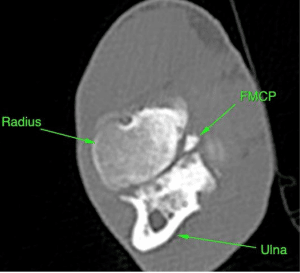
Conditions like fragmented coronoid process usually require CT imaging for accurate diagnosis. CT uses X-rays like traditional radiographs but can provide much more detail and allow a 3D image to be constructed. The detail is required because the area may be small (sometimes only a crack running across the area). We can also assess the incongruency (how well the joint fits together) to some extent.
There are times when CT does not show any changes. In these circumstances we may want to take a sample of the joint fluid (arthrocentesis) for analysis. This can provide useful information about what is happening inside the joint.
Treatment
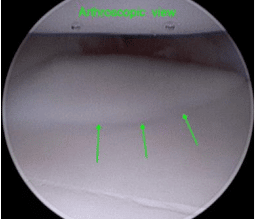
The most common way that we manage a fragmented coronoid process would be to perform a keyhole procedure known as arthroscopy. During this procedure a small camera in inserted into the joint through a small incision in the skin. This allows us to examine the joint, providing more information about the underlying condition. The health of the cartilage can be assessed, which may give an indication about further lameness episodes. Should a fissure or fragment of the coronoid process be present then it is normally removed at this time. If an OCD cartilage flap was present this would also be treated.
The procedure also allows us to decide if additional treatment may be required. Young dogs may benefit from an ulna osteotomy. The ulna is cut below the elbow joint. The aim of this is to improve the congruency of the joint.
Unfortunately many patients with elbow dysplasia will have recurrent lameness episodes. This may be for several reasons but partly will be due to ongoing wear and tear. A dysplastic joint also cannot be made normal. It can be improved but it is always a risk for lameness. There are some ways to reduce ongoing problems which will be discussed during the consultation.