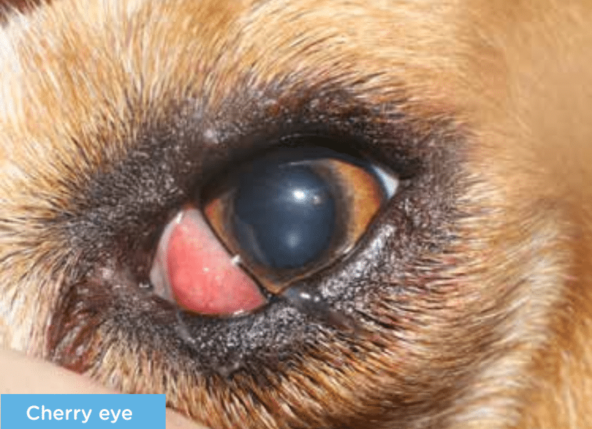
Cherry eye – Prolapse of the gland of the third eyelid
What is the third eyelid gland?
The third eyelid gland is a tear gland located behind the third eyelid and contributes between 30-40% of the total tear film. The tear film is necessary for the supply of oxygen and nourishment to the cornea, providing corneal health and clarity. Without proper function of the third eyelid gland there is a high risk for the development of dry eye disease.
What is Cherry eye and why does it occur?
Cherry eye is the common term for a prolapse of the third eyelid gland. It is called cherry eye due to the characteristic appearance of a pink lump in the inner corner of the eye, resembling a cherry. The exact mechanism why cherry eye occurs remains a mystery. Instead, however, poor attachment of the gland to the underlying tissue as well as a potential genetic component are current theories.
Can both eyes be affected?
Yes, but usually not at the same time. Typically the other eye becomes affected within several months after the first eye.
Are any breeds predisposed?
Whereas cherry eye can affect every breed of dog, it is usually seen in breeds such as English and French Bulldogs, Great Danes, Lhasa Apsos and Shih Tzus, to name a few.
What treatment options do I have?
The recommended treatment for cherry eye is surgery. Whereas there are dozens of surgical treatment methods for treatment of cherry eye, we usually perform a modified version of the so called ‘pocket technique’. We highly discourage surgical removal of the third eyelid gland as it usually results in dry eye disease, requiring life-long treatment.
What is the success-rate of surgical treatment?
The current literature reports a success-rate of 80% however we expect our success-rate to be higher, based on our experience.
What happens if I agree to have the surgery performed?
After a pre-operative physical examination and blood test, surgery is usually performed the same day. Your pet will be discharge either the same day, or the following day, if post-operative monitoring is required. Aftercare includes systemic anti-inflammatory and antibiotic medication as well as antibiotic and lubricating eye drops. The exact treatment plan will be discussed upon discharge. A postoperative check-up is usually scheduled 1 week after surgery and a final check-up at 4 weeks after surgery. Suture removal is not necessary, as absorbable suture material is used.
What happens if I do nothing?
If no surgery is performed, the prolapsed gland will remain in this position. Not only is it cosmetically disturbing and irritating to your pet, but the function of the gland will deteriorate over time, predisposing the eye to the development of dry eye disease, which may require life-long therapy and result in secondary complications (i.e. corneal disease).