Oncology Surgery for Pet Owners
Oncologic (cancer) surgery
Tumours, masses and cancers are terms that are often used interchangeably. Although one sounds more “scary” than the others, they are in essence the same. Tumours are primarily divided into benign and malignant forms. Benign tumours can grow but do not invade or spread. Malignant tumours can invade the surrounding tissue and / or spread to other parts of the body. Typically, a biopsy is needed to determine the difference. A pathologist examines the biopsy under a microscope to gain more information about the tumour, including its origin and how aggressive it is likely to be. This analysis is known as histopathology. Biopsies can be a portion of the larger mass or the whole mass following surgical removal.
The aim of surgery for animals with cancer varies from patient to patient. In some cases, we aim to cure the animal completely. Unfortunately, in others this is not possible, but removing the mass can still be beneficial by making the animal more comfortable or increasing the success of post-operative chemotherapy (medical treatment) or radiotherapy. We always work very closely with our colleagues in the medical oncology service to ensure the best possible outcome for your pet.
Tumours can affect any part of the body including the skin, deeper tissue and organs such as the liver or lungs. Skin masses (including those beneath the skin) are of the most common kind of cancers that we encounter. Like all tumours, they can range from small to large, and benign to aggressive. To better determine the extent of the disease further diagnostic tests are likely to be carried out. This is known as tumour staging. This may include a CT scan, an ultrasound or X-rays. We may also suggest taking a tiny needle sample from the draining lymph nodes (glandular tissue) to determine if there has been any local spread.
Removal of skin tumours can be challenging because we often aim to remove the mass along with a rim of healthy tissue to ensure that all of the cancer cells have been removed. Where this is possible, we must then have a plan to close the wound. This can be achieved via direct closure, skin flaps and skin grafts. In many cases there are several options, which will be discussed with you either at the primary consultation, or after the diagnostic procedures. Many internal cancers can also be removed, even those that are considered very large. This often involves removal of part of the affected organ, for example removal of a portion of the lung or liver (see below).
We have a lot of experience in treating difficult tumours in all parts of the body. We will always
discuss the treatment plan and the prognosis with you in detail before undertaking any
procedure. We work with our Specialist in Anaesthesia and Pain Relief to ensure a suitable
anaesthetic plan incorporating the use of local anaesthetics wherever possible.
It is extremely important that all decisions are made in the best interests of the pet and their
owners. Costs vary depending on the site and size of the tumour, but this will always be discussed
with you at the primary consultation.
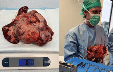
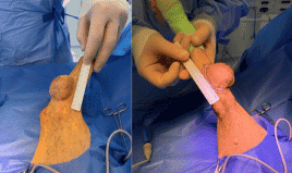
liver mass that was successfully removed from a dog (right). Both of these dogs recovered very well
and were discharged after only one night hospitalization.